Loculated Pleural Effusion Ct Scan / Empyema and Thoracoscopic Surgery, Mumbai, India / Pleural fluid is seen extending to the right oblique fissure.. In patients with loculated pleural fluid the diagnosis of pe had been delayed for a mean of 12.2 days after. Loculated effusion chest ct scan. When the drainage was less than 100 ml/day, urokinase was instilled through the catheter until the drainage was less than 50 ml/day. Us can also guide thoracentesis and placement of tubes into pleural space for drainage. However, ct can help distinguish between a pleural effusion and a pleural empyema (see pleural effusion vs pleural empyema).
The key to resolution involves prompt drainage of pleural fluid because delay leads to the formation of loculated pleural fluid. When the drainage was less than 100 ml/day, urokinase was instilled through the catheter until the drainage was less than 50 ml/day. Debris or septations may be seen in hemothorax or empyema. An effusion was considered loculated when it showed septations, was compartmentalized or accumulated in a fissure or a nondependent portion of the pleura, or when it showed a convex shape facing the lung parenchyma. Conventional chest radiography and computed tomography (ct) scanning are the primary imaging modalities that are used for evaluation of all types of pleural disease, but ultrasound and magnetic resonance.
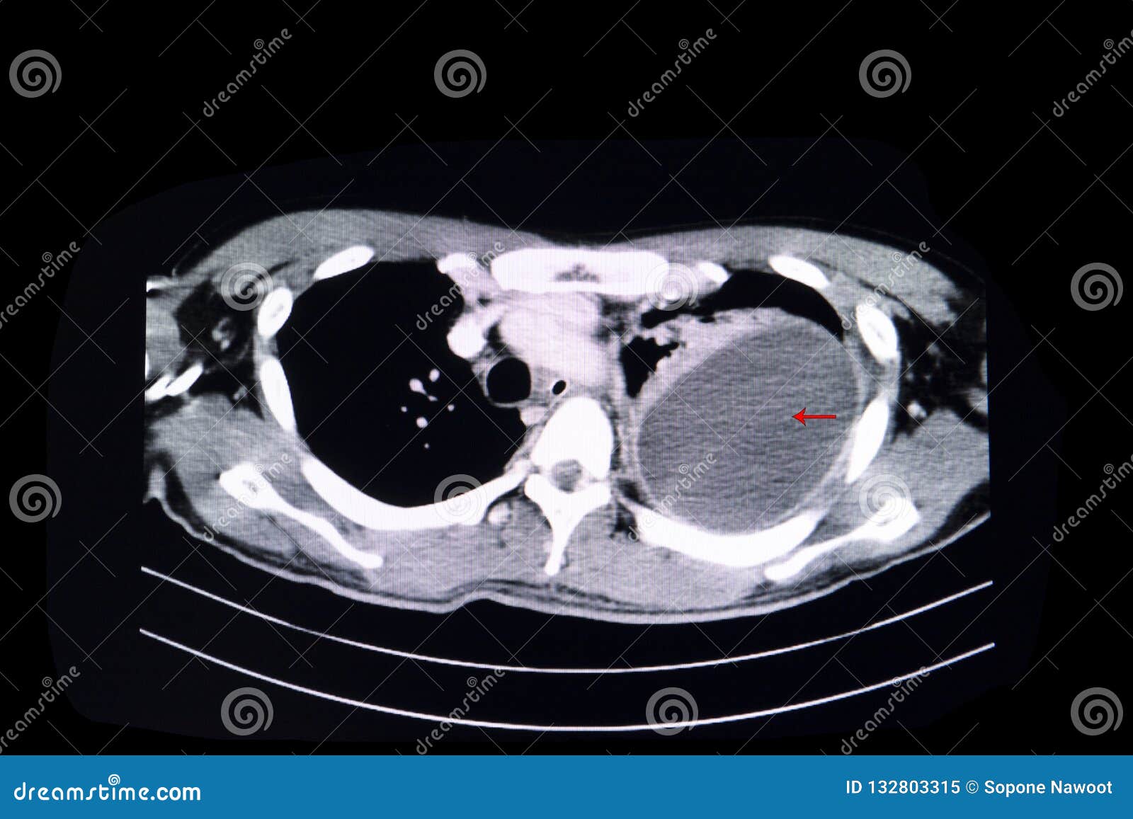
Loculated pleural effusion stock image. Image of computer ... from thumbs.dreamstime.com We develop an automated method to evaluate pleural effusion on ct scans, the measurement of which is prohibitively time consuming when performed manually. Conventional chest radiography and computed tomography (ct) scanning are the primary imaging modalities that are used for evaluation of all types of pleural disease, but ultrasound and magnetic resonance. The key to resolution involves prompt drainage of pleural fluid because delay leads to the formation of loculated pleural fluid. The patient was diagnosed with pneumonia with a loculated parapneumonic effusion. Pleural fluid may loculate within the fissures, particularly in heart failure, and manifest as vanishing or phantom tumors (pseudotumors). Ct scan of thorax shows loculated pleural effusion on left and contrast enhancement of visceral pleura, indicating the etiology is likely an empyema. Us can also guide thoracentesis and placement of tubes into pleural space for drainage. Ct scan of the chest.
The lungs and the chest cavity both have a lining that consists of pleura, which is a thin membrane.
Ideal for localizing, loculated or small effusions for thoracentesis. Ct scan of the chest. The largest pocket of fluid is present posteriorly at the right lung base, with associated atelectasis and minor consolidation. Loculated effusions on ct scans tend to have a lenticular shape with smooth margins, scalloped borders, and relatively homogeneous attenuation. We analyzed ct and sonographic scans of 31 patients with loculated pleural effusion treated with intracavitary urokinase. Ct scan of the chest. We develop an automated method to evaluate pleural effusion on ct scans, the measurement of which is prohibitively time consuming when performed manually. Loculated pleural effusions may be treated by thoracentesis, thoracoscopy, thoracostomy tube drainage, open drainage, or thoracotomy and decortication. Causes of pleural effusion that can be effectively treated or controlled include an infection due to a virus, pneumonia or heart failure. Courtesy of charlie strange, md. Medially situated fluid collections adjacent to the mediastinum are easily delineated on ct scan. Nodular thickening of the pleura suggests pleural metastasis. Axial contrastenhanced ct scan showing moderate left pleural effusion as loculated collection with thickening of pleura (arrows) in a case of mesothelioma open in a separate window figure 14 (a, b)
We analyzed ct and sonographic scans of 31 patients with loculated pleural effusion treated with intracavitary urokinase. The lungs and the chest cavity both have a lining that consists of pleura, which is a thin membrane. Chest ct scan with intravenous contrast in a patient with mixed. Mesothelioma presenting as a pleural effusion: Pleural effusion is an important biomarker for the diagnosis of many diseases.
CT scans of the chest. Left: Computed Tomography scan o ... from openi.nlm.nih.gov Intrapleural urokinase for loculated pleural effusions (pollak, passik) indicated by numerous air bubbles on chest radiography. Depending on the clinical context, ultrasonography or computed tomography (ct) scanning can be used to confirm a pleural effusion, especially in cases of loculated pleural effusion, complete. The patient was diagnosed with pneumonia with a loculated parapneumonic effusion. All ct scans were also reviewed for the presence of additional pleural effusion features. Pleural fluid may loculate within the fissures, particularly in heart failure, and manifest as vanishing or phantom tumors (pseudotumors). We develop an automated method to evaluate pleural effusion on ct scans, the measurement of which is prohibitively time consuming when performed manually. Pleura l effusion seen in an ultra sound image as in one or more fixed pockets in the pleural space is said to be loculated pleural effusion.in us scan us scan they can be identified clearly and it is very complicated.pleural effusion generally found the space. Pleural effusion may extend into the major fissures and give them a characteristic appearance.
Us can also guide thoracentesis and placement of tubes into pleural space for drainage.
The largest pocket of fluid is present posteriorly at the right lung base, with associated atelectasis and minor consolidation. This can be done at the bedside. Pleural tumors, cysts, and loculated effusions all appear homogeneous on chest radiographs, but should be distinguished with ultrasound, ct, or magnetic resonance imaging (mri) scanning. Loculated effusion chest ct scan. We analyzed ct and sonographic scans of 31 patients with loculated pleural effusion treated with intracavitary urokinase. The patient was diagnosed with pneumonia with a loculated parapneumonic effusion. Pleural fluid may loculate within the fissures, particularly in heart failure, and manifest as vanishing or phantom tumors (pseudotumors). Ct of the chest showing a loculated pleural effusion (arrows), which is parapneumonic in origin. Pleural tumors may be regarded as solid, but they are not always entirely homogeneous. Axial contrastenhanced ct scan showing moderate left pleural effusion as loculated collection with thickening of pleura (arrows) in a case of mesothelioma open in a separate window figure 14 (a, b) Pleural effusions are characterized on ct by attenuation values between those of water (0 hounsfield units hu) and soft tissue (approximately 100 hu), typically in the order of 10 to 20 hu. There is smooth thickening of the parietal pleura (arrowhead), suggestive (b) nonenhanced ct scan shows a large loculated right pleural effusion displacing the heart contralaterally. Ct scan of the chest.
Pleural masses may be seen. On ct, 21% of pleural effusions showed loculation. Medially situated fluid collections adjacent to the mediastinum are easily delineated on ct scan. Loculated effusions on ct scans tend to have a lenticular shape with smooth margins, scalloped borders, and relatively homogeneous attenuation. Ideal for localizing, loculated or small effusions for thoracentesis.
Loculated Pleural Effusion Diagram - Chest Radiograph ... from els-jbs-prod-cdn.jbs.elsevierhealth.com Ct of the chest showing a loculated pleural effusion (arrows), which is parapneumonic in origin. Pleural effusions are characterized on ct by attenuation values between those of water (0 hounsfield units hu) and soft tissue (approximately 100 hu), typically in the order of 10 to 20 hu. Chest ct scan with intravenous contrast in a patient with mixed. Loculated pleural effusions may be treated by thoracentesis, thoracoscopy, thoracostomy tube drainage, open drainage, or thoracotomy and decortication. Depending on the clinical context, ultrasonography or computed tomography (ct) scanning can be used to confirm a pleural effusion, especially in cases of loculated pleural effusion, complete. Intrapleural urokinase for loculated pleural effusions (pollak, passik) indicated by numerous air bubbles on chest radiography. All ct scans were also reviewed for the presence of additional pleural effusion features. The patient was diagnosed with pneumonia with a loculated parapneumonic effusion.
Pleural fluid may loculate within the fissures, particularly in heart failure, and manifest as vanishing or phantom tumors (pseudotumors).
Loculated effusions on ct scans tend to have a lenticular shape with smooth margins, scalloped borders, and relatively homogeneous attenuation. Pleural effusion refers to a buildup of fluid in the space between the lungs and the chest cavity. Chest ct scan with intravenous contrast in a patient with mixed. 3 & 4] confirmed the appearance of a complex pleural effusion with associated consolidation of the right hemithorax. Loculated effusion chest ct scan. Pleural fluid may loculate within the fissures, particularly in heart failure, and manifest as vanishing or phantom tumors (pseudotumors). Pleural effusion may extend into the major fissures and give them a characteristic appearance. Pleural effusions are characterized on ct by attenuation values between those of water (0 hounsfield units hu) and soft tissue (approximately 100 hu), typically in the order of 10 to 20 hu. Chest ct revealed a large loculated left pleural effusi. Ideal for localizing, loculated or small effusions for thoracentesis. This can be done at the bedside. Ct of the chest showing a loculated pleural effusion (arrows), which is parapneumonic in origin. Nodular thickening of the pleura suggests pleural metastasis.
Pleural tumors, cysts, and loculated effusions all appear homogeneous on chest radiographs, but should be distinguished with ultrasound, ct, or magnetic resonance imaging (mri) scanning loculated pleural effusion. We analyzed ct and sonographic scans of 31 patients with loculated pleural effusion treated with intracavitary urokinase.


0 Komentar